
Ankylosing Spondylitis Radiology Key
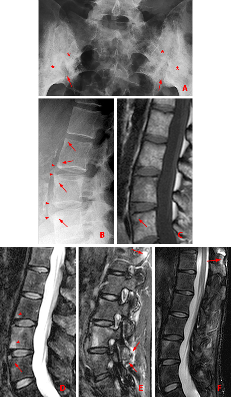
The earliest signs of spondylitis are manifest as small erosions at the corners of the vertebral bodies - the so-called Romanus lesion. Syndesmophyte formation eventually lead to classical 'bamboo spine'. Osteoporosis and kyphosis occur with long-standing disease. Extra-axial skeletal involvement mimics mild rheumatoid arthritis.
Ankylosing Spondylitis Radiology Key
Imaging in ankylosing spondylitis (AS) has been synonymous for decades with conventional radiography (CR). However, developments in computed tomography (CT), ultrasonography (US) and particularly magnetic resonance imaging (MRI) have dramatically increased the amount and scope of information obtainable by imaging.

Image
Ankylosing spondylitis (less commonly known as Bechterew disease or Marie Strümpell disease ) is a seronegative spondyloarthropathy , which results in fusion (ankylosis) of the spine and sacroiliac (SI) joints, although involvement is also seen in large and small joints. Epidemiology

Ankylosing spondylitis lumbar spine Image
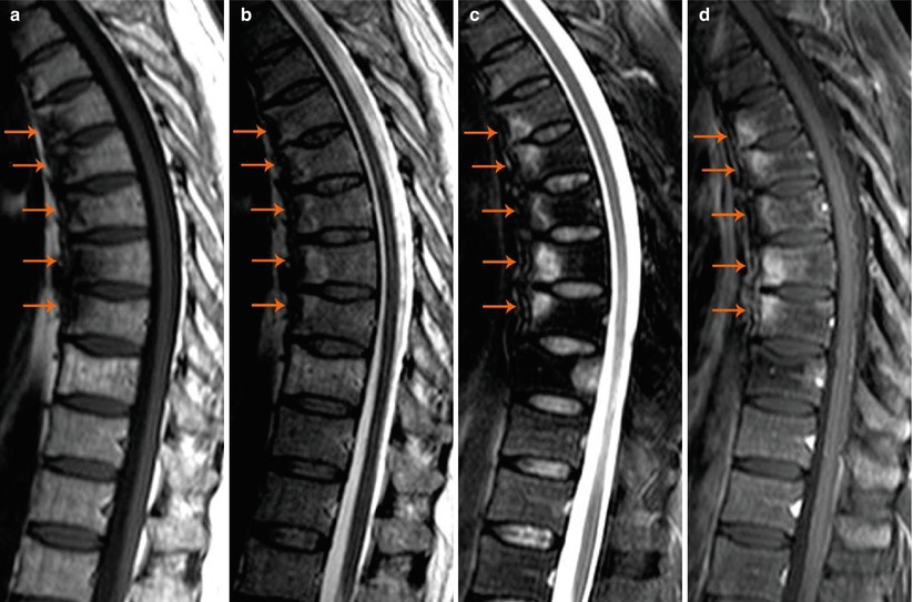
Ankylosing spondylitis (AS) is a chronic inflammatory disease affecting the spine and the sacroiliac joints. AS occurs with the inflammation of the entheses and formation of syndesmophytes and finally sacral and spinal ankylosis. Imaging demonstrates both inflammatory and chronic lesions. Sacroiliitis is the hallmark of the disease. Spinal changes usually take place in advanced stages of the.

Ankylosing Spondylitis Mri
Ankylosing spondylitis (AS) is a chronic inflammatory disease that primarily affects the spinal and sacroiliac joints, which link the pelvis and spine. Fusion (ankylosis) of the spine's vertebrae, which happens over time, is a hallmark sign of this disease.

Ankylosing spondylitis Radiology Case Ankylosing spondylitis, Radiology, Case
Objective: To update evidence-based recommendations for the treatment of patients with ankylosing spondylitis (AS) and nonradiographic axial spondyloarthritis (SpA). Methods: We conducted updated systematic literature reviews for 20 clinical questions on pharmacologic treatment addressed in the 2015 guidelines, and for 26 new questions on pharmacologic treatment, treat-to-target strategy, and.

Ankylosing spondylitis "Romanus lesions" Image
Abstract. Imaging has a central role in the diagnosis, management, and follow-up of patients with axial spondyloarthritis (axSpA). For the early diagnosis of axSpA, magnetic resonance imaging is of utmost relevance. While no novel imaging techniques were developed during the past decade, improvements to the existing modalities have been introduced.
Ankylosing Spondylitis Radsource
Ankylosing spondylitis (AS) is a chronic inflammatory rheumatologic disorder that predominantly affects the axial skeleton and is characterized by sacroiliitis, spondylitis and enthesitis.
Ankylosing Spondylitis and How It Is Portrayed Radiographically
Clinical Presentation The patient is a 65-year-old female with a history of chronic low back and neck pain and markedly reduced mobility in the cervical and lumbar region. Her records have noted the diagnosis of ankylosing spondylitis for 20 years.

Image
CT scan X-ray Treatment The goal of treatment is to relieve pain and stiffness and prevent or delay complications and spinal deformity. Ankylosing spondylitis treatment is most successful before the disease causes irreversible damage. Medications

Imaging in ankylosing spondylitis Mikkel Østergaard, Robert G.W. Lambert, 2012
Case Discussion. Ankylosing spondylitis is a chronic inflammatory disease of the spine characterized by Chronic back pain and progressive spinal stiffness. Involvement of the spine, sacroiliac joints, peripheral joints, digits, and entheses are characteristic.

Andersson lesion in ankylosing spondylitis BMJ Case Reports
Imaging is an integral part of the management of patients with ankylosing spondylitis and axial spondyloarthritis. Characteristic radiographic and/or magnetic resonance imaging (MRI) findings are key in the diagnosis. Radiography and MRI are also useful in monitoring the disease.

Images
Ankylosing spondylitis is the most common seronegative spondyloarthropathy. It has characteristic radiological features.

Ankylosing spondylitis. Xray AP (a) and lateral (b) lumbar spine.... Download Scientific Diagram
Preferred examination Radiographs are the single most important imaging technique for the detection, diagnosis, and follow-up monitoring of patients with ankylosing spondylitis. Overall bony.
Ankylosing Spondylitis MRI Sumer's Radiology Blog
Patterns of radiographic involvement can be assessed using the Bath Ankylosing Spondylitis Radiology Index (BASRI). Usually, symmetric sacroiliitis can be seen in 86% of patients, complete spinal fusion in 28% of patients for more than 30 years, and in 43% of patients with AS for more than 40 years. 13

Spinal injury in ankylosing spondylitis The BMJ
Spondylosis is a seronegative spondyloarthropathy typically seen in young males affecting the sacroiliac joint first with multisystemic manifestations and multiple associations.
